Women Should Know Their Breast Density: Here’s Why

More than half of women over 40 in the United States have dense breasts. Many of them don’t know it.
[Video is in Spanish.]
But starting in September 2024, all mammogram reports sent to patients in the U.S. will be required to include information about breast density.
“Here at Sylvester and in Florida we have been communicating these results to patients for approximately 10 years, but the Food and Drug Administration (FDA) standardized this requirement at the national level,” says Monica Yepes, M.D., a radiologist specializing in breast imaging from Sylvester Comprehensive Cancer Center, part of the University of Miami UHealth System.
Mammography is the only way a woman can find out her breast density.
This communication must explain what breast density consists of, whether the patient has dense tissues or not, and why breast density is a risk for breast cancer.
The breasts are made up of fatty breast tissue and dense breast tissue. The dense tissue includes the mammary ducts and lobes, which is where milk is produced and transported. “People who have more dense tissue have a greater number of ductal and lobular structures, so they have a greater predisposition to develop breast cancer”, explains Dr. Yepes.
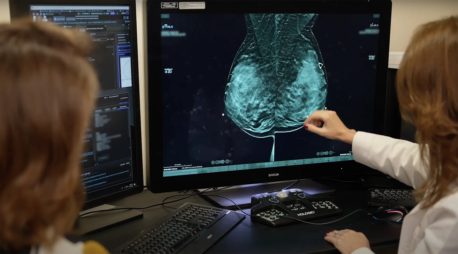
Breast density can affect the appearance of breast tissues on a mammogram and mask cancer. “The problem is that dense tissue looks white on mammography and cancers are white too,” says Dr. Yepes. “In non-dense tissue, the cancer protrudes very well, while in dense white tissue, it does not protrude,” she adds.
After a mammogram, women with dense breasts may need additional testing to increase the chance of finding cancer.
“These studies can range from ultrasound, which is what we usually recommend for women who are not at high risk, and if they are at very high risk, we could recommend an MRI or a mammogram with contrast”, says the radiologist.
Mammography with contrast medium is very useful for making an early diagnosis and, especially, in women with dense tissues and at high risk of breast cancer. “The contrast goes to the sites where there is increased vascularization. Cancers form vascularization and the contrast medium accumulates abnormally there”, explains Dr. Yepes.
At Sylvester’s Breast Imaging Department, all radiologists specialize in breast cancer diagnosis and have the experience necessary to produce accurate reports.
“It has been shown that when patients go to an oncologist designated by the National Cancer Institute (NCI), they will be evaluated by experts who have a greater probability of finding cancer and will have a greater survival if they develop cancer”, he says. Dr. Yepes.
Sylvester has a program for women at high risk of breast cancer. “Based on the risk factors that a patient has, such as family history, the presence of genetic mutations and a previous biopsy that has given an atypical result, we can calculate the patient’s risk of developing cancer and perform a recommendation for further study.”
For more information about the program for women at high risk of breast cancer, call 305-689-7475 or visit our cancer prevention website.
Article and video written and produced by Shirley Ravachi for ‘Cuidando Su Salud’, a series of healthcare-related stories that air regularly on Telemundo 51. For more stories like this, visit the Sylvester Comprehensive Cancer Center YouTube channel.
Tags: Breast Cancer, Breast Density, Breast Imaging, mammogram, MD., Monica Yepes, radiology, Sylvester Comprehensive Cancer Center
