
You Got a Mammogram. Now What?
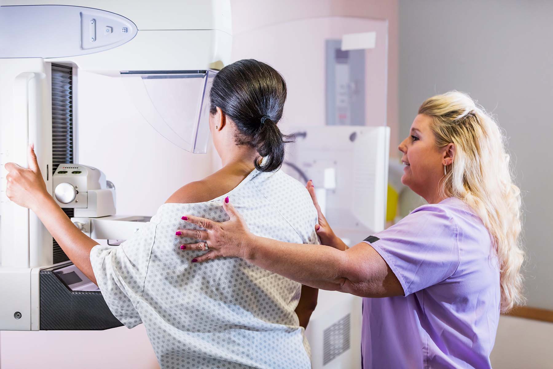
You made the appointment, showed up at the breast imaging center and endured the uncomfortable compression. Now comes the part many women find surprisingly stressful.
Waiting for the results.
What happens next depends on what the radiologist sees on your mammogram and, sometimes, on what has changed since your previous images.
For most women, the next step is simply to have a conversation with a medical care provider or receive a report showing that no suspicious abnormality was found, then return for their next routine screening. But sometimes the imaging center asks you to come back for additional images. That can sound alarming, but a callback does not mean you have breast cancer.
Here’s what to know about what happens after a mammogram.
First: A radiologist reviews your mammogram images
A radiologist who specializes in breast imaging examines the images from your mammogram, often comparing them with images from previous years. This crucial comparison can help distinguish a normal variation from something that is new or has changed.
Your report will include a standardized assessment called a BI-RADS category. BI-RADS, short for Breast Imaging Reporting and Data System, helps radiologists communicate what they see and what follow-up, if any, is recommended.
BI-RADS categories for screening mammograms include:
- Negative: No findings. Stick to routine screening.
- Benign: Something was seen, but it is definitely non-cancerous (such as a cyst). Sttick to routine screening.
- Incomplete, additional imaging needed: You’ll get a callback for a closer look.
Second: You might get a callback
A callback is not a diagnosis of cancer. It just means the radiologist wants a closer look at something seen on the screening mammogram. Often, a better or different image is needed to determine whether an area represents normal breast tissue. Keep in mind that less than 10% of women called back for more imaging are found to have cancer.
For your callback, you might be asked to have another mammogram with additional views, known as a diagnostic mammogram. You might also be asked to have a breast ultrasound, which gives a different view of the breast tissue.
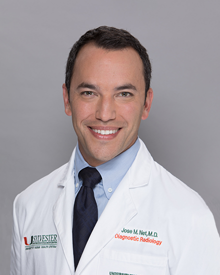
“In our practice, around 15% of women will be called back for an additional workup,” says Jose Net, M.D., director of breast imaging services at Sylvester Comprehensive Cancer Center, part of UHealth—University of Miami Health System. “The majority of these workups answer any questions we have and don’t lead to a cancer diagnosis. Most women return to routine screening afterward.”
Try not to interpret a callback before the additional testing is complete. The purpose of diagnostic imaging is to gather more information.
You might see a note about dense breasts
You may also see the words “dense breast tissue” on your mammogram report.
Breast density refers to how much fibrous and glandular tissue you have compared with fatty tissue. It is a normal characteristic of the breast, not a disease, and it can change over time. Almost half of all women over 40 who get a mammogram have dense breast tissue.
Dense breasts can make some cancers harder to see on a mammogram, and having dense breasts is associated with a higher risk of breast cancer.
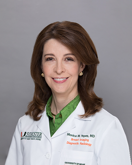
“Breast density is one of the most common questions patients have after receiving a mammogram report,” says Monica Yepes, M.D., a Sylvester radiologist specializing in breast imaging. “Dense breast tissue is normal, but it can affect how we interpret imaging and may influence whether additional screening tests, such as an ultrasound, MRI or contrast-enhanced mammogram, should be considered, based on a woman’s overall risk profile.”
If your mammogram shows dense breasts, talk with your health care provider about what that means for you and whether additional screening makes sense based on your overall risk.
When is a breast biopsy needed?
If additional imaging shows an area that remains concerning, your doctor may recommend a biopsy. This does not mean that you have cancer.
A biopsy is the only way to determine whether certain abnormal breast tissue is cancerous. During a breast biopsy, a doctor removes a small sample of tissue so it can be examined by a pathologist.
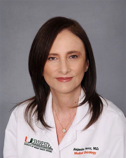
“Most patients who are called back for a biopsy receive benign results,” says Alejandra T. Perez, M.D., a Sylvester breast medical oncologist and leader of Sylvester’s Breast Cancer Assessment Risk Evaluation Clinic. “Nonetheless, if a biopsy has been recommended, it’s critical that you schedule it in a timely fashion. This supports early detection and improves outcomes.”
Hearing the word “biopsy” can understandably be frightening. But needing a biopsy still does not necessarily mean you have cancer. The biopsy is what provides the definitive information needed to make a diagnosis.
Don’t ignore symptoms, even if your last mammogram was normal
A normal mammogram is reassuring, but it does not mean you can ignore a new breast change.
Tell your health care provider if you notice a new lump, nipple changes, skin changes, unusual nipple discharge or another change that concerns you. Screening mammography is designed for people without breast symptoms. A new symptom may require diagnostic evaluation even if your most recent screening mammogram was normal.
Breast cancer screening is an ongoing process.
Breast cancer screening works best when it is part of an ongoing process, rather than a once-a-year event. For women at average risk, the American College of Radiology and Society of Breast Imaging recommends screening mammography every year beginning at age 40. When to start depends on one’s personal risk profile, so your personal screening plan should take your age, risk factors, family history and preferences into account and should be discussed with your health care provider.
And if your mammogram leads to additional testing, remember that more testing is not the same thing as a cancer diagnosis. The goal is to understand what your images show and, when something needs a closer look, to find out what it is.
“Annual screening mammograms are the only intervention proven to reduce death from breast cancer,” Dr. Net notes. “They offer the best chance of detecting breast cancer at its earliest stage, when it is most treatable.”
So, when you leave your next mammogram, don’t just ask, “When will I get my results?”
Ask “What happens next?”
Knowing the answer can make the waiting and whatever comes after a little less uncertain.
Reviewed by Jose Net, M.D., Monica Yepes, M.D. and Alejandra T. Perez, M.D.
Tags: breast cancer early detection, breast cancer risk factors, breast cancer screening, early-stage breast cancer, family history of breast cancer, genetic testing, high-risk breast cancer screening, mammogram, personalized breast cancer screening, women's health

