
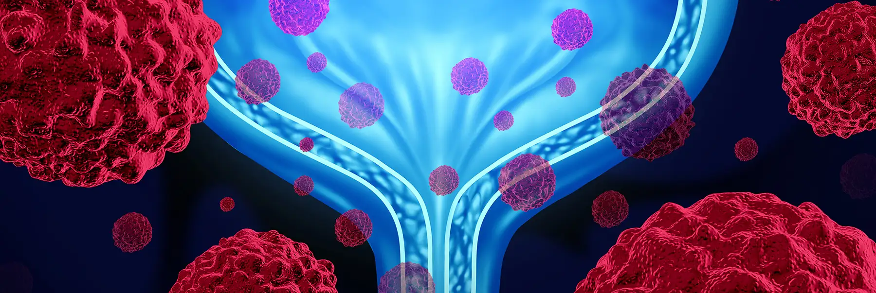
Can AI Improve Bladder Cancer Screening?
Machine-learning and artificial intelligence (AI) are changing how we live and work.
So, can the same technology make diagnostic tests for bladder cancer more accurate and comfortable? An initial research study shows promise.
A new technique for identifying cancerous cells in patients had a 94 percent diagnostic accuracy according to one study. The technology, called atomic force microscopy (AFM), uses machine-learning to analyze images created from cells obtained from a urine sample.
Cystoscopy is the gold standard for bladder cancer diagnosis. Experts insert a cystoscope, a thin tubular instrument, into the urethra and then guide it slowly into the bladder. A tiny video camera at the tip of the scope helps your surgeon see any abnormalities.
“The results suggest a possibly improved way to detect bladder cancer cells compared to cystoscopy and other non-invasive tests,” says Dr. Chad R. Ritch, a urologic oncologist at Sylvester Comprehensive Cancer Center, part of the University of Miami Health System. “However, the research did not compare AFM head to head with cystoscopy. It is too soon to say that it is superior to cystoscopy. Future studies will test that hypothesis.”
How do they screen for bladder cancer now?
“Currently, we diagnose by visualizing the bladder and sampling abnormal areas to microscopically inspect inside the cells,” shares Dr. Ritch. His work focuses on studying predictive biomarkers for bladder cancer.
“This newer possibility looks at something more basic: exterior shapes. The technical term for this analysis is nanoscale mapping. The computer program is analyzing very tiny maps of the exterior cell walls. The cancerous cells had different outlines from the healthy cells.”
How it works
First, specialists gather cells from a patient’s urine sample instead of probing the inside of the bladder. After that, they use the AFM to point a safe laser light at the cells and create images based on mathematical calculations. Then, a computer program uses the power of mathematical learning to compare the shapes and patterns of healthy bladder cells.
“This is a first trial of this approach,” says Dr. Ritch. “The researchers looked at cells from patients already being treated for bladder cancer versus cells from those without the disease.”
Benefits beyond bladder cancer?
If follow-up studies show the same outcomes, then the world may one day have a far less invasive, and more accurate, way to diagnose bladder and additional cancers. Dr. Ritch says that the test would have great value for anyone already in treatment for bladder cancer.
“After you are diagnosed with bladder cancer, treated and cancer-free, you still need follow-up cystoscopies,” he says. “We need to ensure the cancer does not come back. This new approach would make repeated follow-ups much more comfortable and convenient for patients at every stage of their disease. It could potentially allow for fewer cystoscopies.”
Learn more about bladder cancer today.
John Senall is a contributing writer for UMiami Health News. He is a former hospital and comprehensive cancer center communications director.
Tags: artificial intelligence, Atomic force microscopy, bladder cancer, diagnostics, Dr. Chad Ritch, Sylvester Comprehensive Cancer Center