
Artificial Intelligence: A Game Changer in Brain Tumor Surgery?
Thanks to new cutting-edge technology, neurosurgeons can diagnose brain tumors during surgery, according to a recent study.*
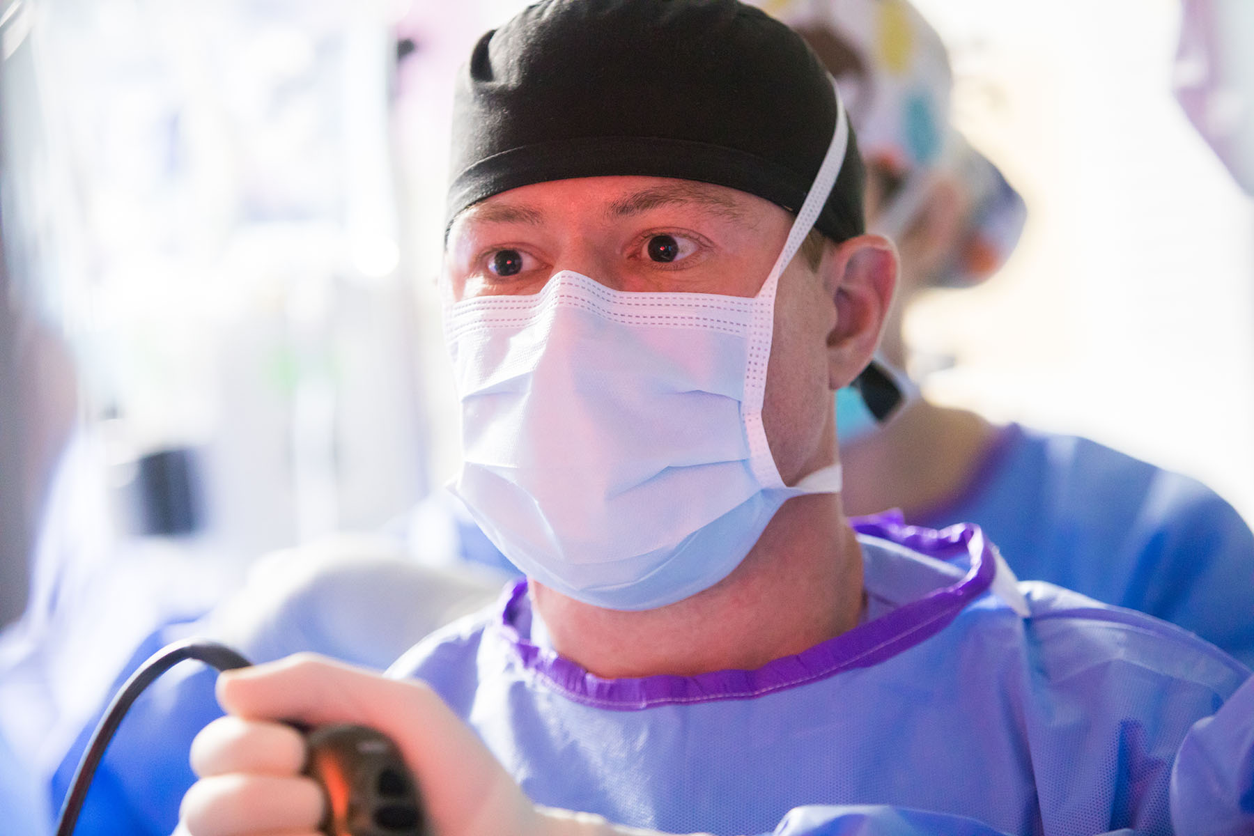
“It’s really a step forward in providing rapid intraoperative [during surgery] diagnoses of malignant and benign tumors, which is essential information needed to make critical decisions during safe and effective brain tumor surgery,” says Sylvester neurosurgeon and study co-author Dr. Michael Ivan, M.B.S., who played a major role in developing the Stimulated Raman Histology (SRH) technology with leading collaborators at New York University and the University of Michigan.
An accurate diagnosis is crucial during the actual surgical removal of brain tumors. T
he technology uses artificial intelligence (AI) along with optical imaging for a near real-time reading. “This digitized process provides surgical teams a diagnosis in less than three minutes as opposed to a 20-30 minute wait time during a traditional process.”
Conventional methods for intraoperative histology, or the study of tissues, are time-consuming and require numerous steps – each of which represents a potential barrier to delivering safe, timely, and effective surgical care. These include sectioning and freezing of the tissues, tissue transport to a pathology laboratory, specimen processing, slide preparation by highly trained technicians, and interpretation by pathologists.
The study examined the accuracy of brain tumor image classification through machine learning and compared it with the precision of conventional methods. The results for both were comparable: the AI-based diagnosis was 94.6% accurate, compared with 93.9% for the pathologist-based interpretation on a frozen specimen.
One of the most significant advantages? SRH ensures more precision in completely removing cancerous brain tumors, says Dr. Ivan, who is a leader of neuro-oncology at Sylvester and assistant professor of neurological surgery at the Miller School of Medicine.
In many of our surgeries on malignant tumors, the ability to remove all of the tumor makes a difference in a patient’s overall survival.
Dr. Michael Ivan
“Artificial intelligence provides more rapid and frequent information to the surgeon while operating to ensure the boundaries of the surgical resection are clear of cancer.”
This game-changing technology is an exciting step forward in the management of brain tumors, says Dr. Ricardo Komotar, a study co-author and director of surgical neuro-oncology and Sylvester’s Brain Tumor Initiative.
How does Stimulated Raman Histology work?
The ground-breaking imaging technique shows any tumor infiltration in human tissue. By collecting scattered laser light, the imaging illuminates essential features not typically seen in standard images of the tissues.
The microscopic images are then processed and analyzed with AI. In under two and a half minutes, surgeons can see a predicted brain tumor diagnosis. Using the same technology, after the resection, they were also able to accurately detect and remove an otherwise undetectable tumor.
“This is a very complex AI system, which looks at patterns, intensity, and coloration of the specimen’s digital image to provide an instantaneous diagnosis,” says Dr. Ivan.
How the study was conducted
To build the tool, researchers designed an AI model trained on a deep convolutional neural network (CNN) that includes more than 2.5 million images taken from the samples of 415 patients. The AI uses CNN to classify tissue into 13 categories that represent the most common brain tumors, including malignant glioma, lymphoma, metastatic tumors, and meningioma.
Dr. Ivan and researchers and neurosurgeons at two other sites enrolled 278 patients undergoing brain tumor resection or epilepsy surgery in the clinical study. Brain tumor specimens were biopsied from patients, split intraoperatively into sister specimens, and randomly assigned to the control or experimental arm.
Specimens routed through the control group — the current standard practice — were transported to a pathology laboratory and went through conventional specimen processing. The experimental group was performed during surgery, from image acquisition and processing to diagnostic prediction via CNN.
The study showed that the diagnostic errors in the experimental group were different from the errors in the control group, suggesting that a pathologist using the novel technique could achieve close to 100% accuracy. The system’s precise diagnostic capacity could benefit centers that don’t have access to expert neuropathologists.
“As surgeons, we’re limited to acting on what we can see; this technology allows us to see what would otherwise be invisible, to improve speed and accuracy in the OR, and reduce the risk of misdiagnosis,” said senior author Dr. Daniel A. Orringer, associate professor of neurosurgery at NYU Grossman School of Medicine, who helped develop SRH and co-led the study with colleagues at the University of Michigan. “With this imaging technology, cancer operations are safer and more effective than ever before.”
In addition to improving the workflow of pathologists, researchers say that the technology can be used in other medical settings that also depend on the expert analysis of tumor samples obtained during surgery.
“The combination of artificial intelligence and surgical experience is an example of what separates Sylvester Comprehensive Cancer Center from other centers in the state of Florida,” says Dr. Komotar.
*Study Title: Near Real-time Intraoperative Brain Tumor Diagnosis Using Stimulated Raman Histology and Deep Neural Networks
The collaborative study, co-authored by neurosurgeons with Sylvester Comprehensive Cancer Center, part of the University of Miami Miller School of Medicine, was published on January 6 in the journal Nature Medicine.
Originally written by Kai Hill, contributor for Inventum.
Changes medically reviewed by Dr. Michael Ivan and Dr. Ricardo Komotar.
Tags: brain health, brain tumor care in Miami, Dr. Michael Ivan, Dr. Ricardo J. Komotar